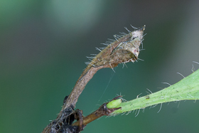
Abstract: As part of a broader study into the life cycle of the White Admiral butterfly, the author has examined the habits of the overwintering larva. In this article he presents four different types of hibernacula that he has encountered so that it may help those looking to monitor them over the winter months.
I am fortunate to live close to Pamber Forest in north east Hampshire, which is one of the best sites for White Admiral in the British Isles. Over the course of both 2015 and 2016 I have taken a particular interest in the immature stages of this species.
The White Admiral overwinters as a 3rd instar larva, hidden inside a hibernaculum that is formed from a single Honeysuckle (Lonicera periclymenum) leaf, which is often the leaf on which the egg was laid. The plant is deciduous and loses its leaves over the winter, and so the larva attaches the leaf stalk to the stem using a number of silk strands so that it does not detach from the plant. A larva will ultimately emerge from its hibernaculum in the spring and feed on the tender leaves of new Honeysuckle growth.
Most hibernacula are ultimately attached to the stem only by these silk strands as the leaf dies and the leaf stalk breaks away from the stem, and I am grateful to Matthew Oates for showing me a technique for finding hibernacula that embraces this characteristic: gently move or blow on the plant to see if any of the leaves sway from side to side (thereby indicating a potential hibernaculum that is held on to the plant only with silk). Any other leaves that are present do not tend to move in this manner, and are usually those that were the last to die and their stalk is still attached to the stem, or they have yet to feel the full force of a winter storm, after which they are normally blown from the plant.
During the course of my study I have encountered 4 types of hibernacula and they are each given a name as described below.
In this type of hibernaculum, the larva deliberately cuts the leaf so that the half furthest from the stem falls away. The larva then lays down silk strands that span each side of what remains of the leaf, causing the sides to come together as the silk dries and contracts. The larva remains in the resulting abode and eventually seals the open ends with silk.
The incredible sequence of shots below have been generously contributed by Wolfgang Wagner who runs the excellent website "Lepidoptera and their ecology" (Lepidoptera and Their Ecology, 2016), from which these photos have been obtained.
The end result of this type of hibernaculum is provided in Figure 12, showing a hibernaculum in its final state which, in this case, is completely sealed using an overlapping flap, although not all examples are so comprehensively sealed in this way.
 |
| Figure 12 - The 'cut and fold' type of hibernaculum (Pamber Forest, 23rd September 2015) Image © Peter Eeles |
This is one of the commonest types of hibernacula that I have come across, accounting for 29.2% of those recorded, from a sample of 48.
This type of hibernaculum is so named because it is the start of the "Cut and Fold" type, but only half of the leaf is cut, and this half is then folded over with silk, creating a chamber within which the larva overwinters.
This type also caters for situations where a larva partially cuts any portion of the leaf (and not always as neatly as shown in Figures 14 and 15). Examples are given in Figure 16, where the cut is not at the base of the leaf but closer to the tip, and in Figure 17, where the leaf has been partially cut at two points. Overall, this type was the most common and accounted for 45.8% of those recorded.
The third type of hibernaculum is where the larva silks (quite neatly) most of the edges of the leaf together, leaving only the ends free of silk. The leaf is not cut, but simply allowed to wither. This is the least common type of hibernaculum, accounting for just 8.3% of those recorded. Figures 18, 19 and 20 are of the same hibernaculum.
The fourth type of hibernaculum is the simplest of all where the leaf is simply "folded" on itself and the two parts silked together. The end result is often quite messy and certainly not as symmetric as the previous type. This type was not very common, accounting for just 16.7% of hibernacula recorded.
Most authors would appear to make simplifying remarks concerning the White Admiral hibernaculum, many of them simply saying that it looks like a shrivelled leaf or assuming that there is only one type of hibernaculum. However, I believe that recognising all types of hibernacula not only provides a more correct interpretation, but may also act as an aid to those of us that search out and monitor White Admiral larvae over the winter.
My particular thanks go to Graham Dennis, Reserves Officer at Hampshire and Isle of Wight Wildlife Trust, for showing me some of the favoured egg-laying spots for White Admiral in Pamber Forest. I would also like to thank Matthew Oates of the National Trust for sharing his experience in locating White Admiral hibernacula and also Wolfgang Wagner for generously donating a series of images that characterise the "cut and fold" type of hibernaculum. Finally, I would like to thank Mark Colvin for providing constructive feedback when reviewing this article.